
8 Reasons to Integrate 3D Printing in Hospitals
3D printing is no longer limited to prototypes, school projects, or industrial parts. Hospitals are using it to create patient-specific anatomical models, surgical guides, prostheses, casts, training models, and other medical tools.
The main advantage is simple. A hospital can turn a patient's CT or MRI data into a physical 3D object that doctors can examine, measure, and use for planning. Here are eight important reasons hospitals are integrating 3D printing into clinical workflows.
1. 3D Printing Makes Patient Anatomy Easier to Understand
Radiology, computed tomography, magnetic resonance imaging, and other imaging modalities are already in use by medical professionals. However, the typical viewing of these images is as a succession of 2D slices. It might be challenging to grasp complex anatomy when many organs, blood arteries, bones, and tumors are in close proximity to one another.
This data becomes a tangible tool for clinicians when they use a 3D-printed model. They may view the interrelationships of structures, spin it, and look at it from all directions before treatment starts.
Real-World Case: Mayo Clinic's Conjoined Twins
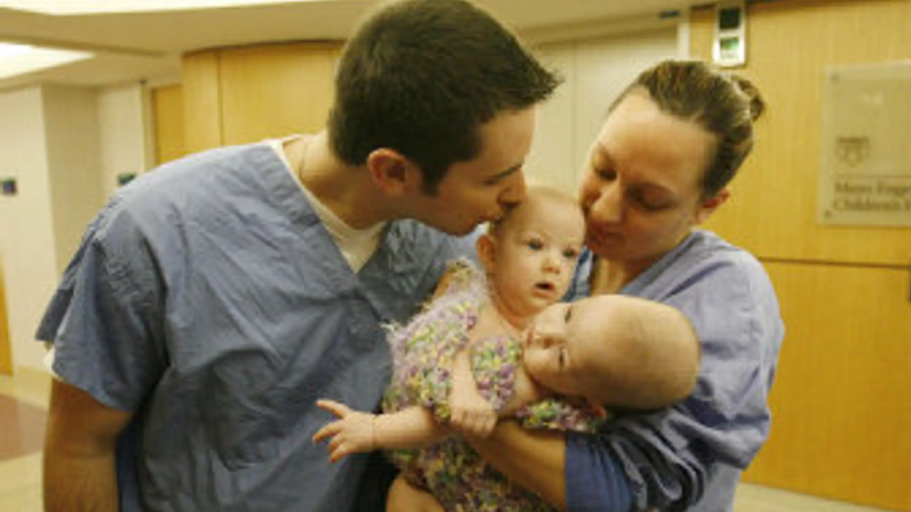
Conjoined twins were the initial complicated case that sparked the Mayo Clinic's 3D printing program. Surgeons had to learn the interconnections of the kids' organs before they could try to separate them. In order to aid surgeons in their understanding of the anatomy, the researchers built a physical 3D model of the shared liver.
That preliminary work laid the groundwork for the current anatomic modeling program at the Mayo Clinic. Cardiovascular surgery, orthopedic surgery, neurosurgery, and cancer treatment are just a few of the areas where the hospital now employs patient-specific models.
2. It Helps Surgeons Plan Complex Operations
Before going into surgery, a surgeon can examine the patient's internal structures. When dealing with abnormally shaped or located tumors, blood vessels, bones, or organs, this can be extremely helpful.
When discussing the same anatomy, a physical model might also be helpful for the surgical team. Experts can collaborate on a single patient-specific model instead of glancing at individual screens.
Real-World Case: A Pancoast Tumor at Mayo Clinic
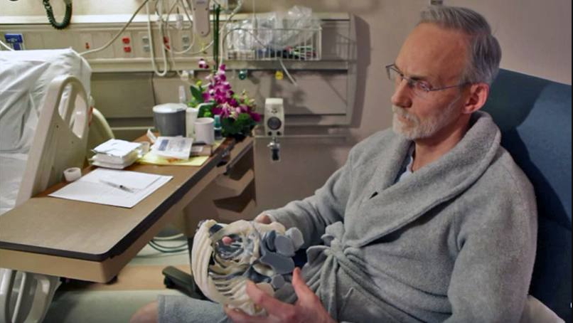
Mayo Clinic employed a 3D-printed model to help plan the excision of a Pancoast tumor located toward the top of a patient's lung. The model highlighted the tumor's interaction with vital neurons and blood arteries. This assisted the surgical team in comprehending the anatomy before the operation.
Mayo Clinic claims its current work includes 3D models for lung, heart, brain, joint, head and neck, and tumor surgery. Its 2025 guidance also includes printing patient-specific models and cutting guides in-house for difficult head and neck cancer operations.
3. Hospitals Can Create Patient-Specific Surgical Guides
Not every patient has the same anatomy. This can make standard surgical tools less suitable for some complex procedures.
3D printing lets surgeons design a guide from the patient's imaging data. The guide can match a specific bone or anatomical structure and help the surgeon follow a planned cutting or drilling path.
Real-World Case: Mayo Clinic Head and Neck Surgery

Mayo Clinic uses virtual surgical planning and 3D-printed cutting guides for some head and neck procedures. Its surgeons describe using patient-specific guides to plan bone cuts and reconstruction.
According to Mayo Clinic, this approach can save up to two hours in some operating-room procedures. The clinic also reports using the models and guides to improve planning for complex head and neck reconstruction.
4. 3D Printing Supports Personalized Implants and Prostheses
Traditional medical implants often come in standard sizes. But some patients have bone defects that are too unusual for standard parts.
3D printing can produce an implant based on the patient's anatomy. It can also create complex shapes and internal structures that are difficult to manufacture with traditional methods.
Real-World Case: A 3D-Printed Scaphoid Prosthesis
Researchers at Cantonal Hospital Baselland and collaborating institutions reported an in-hospital 3D-printed scaphoid prosthesis made from medical-grade PEEK.
The team used an Apium M220 printer designed for medical PEEK applications. The patient-specific prosthesis was produced using fused filament fabrication. The study demonstrated the potential for point-of-care manufacturing of patient-specific bone replacements, although such applications require strict medical and regulatory controls.
5. It Can Help With Custom Orthopedic Devices
Casts, splints, and other prosthetics can be custom-made for each patient using 3D printing technology. The patient's anatomy can be more closely followed by a personalized design than by a conventional product. This has the potential to make the gadget more comfortable and lightweight without sacrificing support.
Real-World Case: 3D-Printed Wrist and Hand Casts
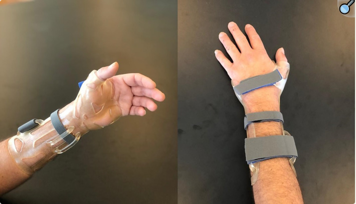
Individualized 3D-printed casts were the subject of an in-hospital workflow evaluation in a clinical trial involving patients suffering from non-displaced fractures of the hand and wrist.
Twenty patients were part of the investigation. On average, the researchers found that the full printing process took around 161 minutes. At the last follow-up, every patient had healed their fractures, and no one had complained of pressure sores or reduction loss. Casting these fractures became standard practice at the medical center.
This demonstrates the potential for 3D printing to enter the realm of direct patient care, expanding its use beyond surgical planning.
6. It Gives Doctors a Better Way to Practice Rare Procedures
Some surgical procedures are rarely performed. A surgeon may therefore have limited opportunities to practice a particular case before treating a real patient.
A 3D-printed model can provide a physical training platform. It can be based on real anatomy or designed to reproduce a difficult medical condition.
Real-World Case: Johns Hopkins Surgical Model
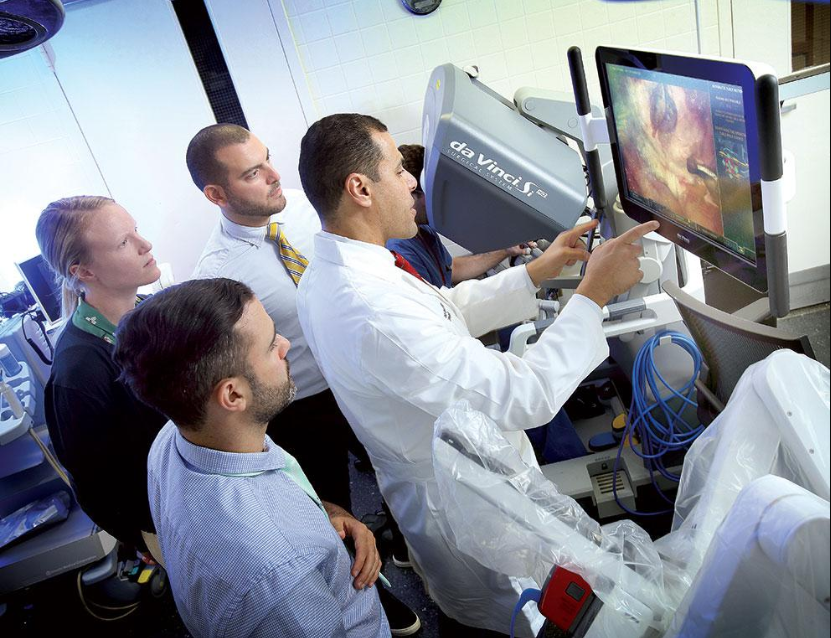
Johns Hopkins uses patient-specific surgical models through its SLICE program. The models are created from individual patient scans and can reproduce anatomy and pathology for practice.
Surgeons can use these models to plan and modify their approach before the actual operation. Johns Hopkins says its models are intended to reduce uncertainty in complex and rare surgical cases.
This is particularly useful when the normal learning process would otherwise depend on encountering a similar case during surgery.
7. 3D-Printed Models Improve Medical Education
Medical students normally learn anatomy from textbooks, digital images, cadavers, and plastic models. These tools remain important, but 3D printing adds another option.
A hospital can print a specific heart, bone, tumor, blood vessel, or other structure. Students can then study the shape and relationships of the anatomy from multiple angles.
Real-World Case: Mayo Clinic's Pediatric Neurosurgery Models
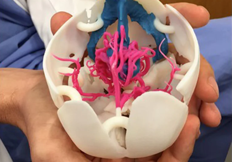
Mayo Clinic created 3D-printed models of conditions such as aneurysms, arteriovenous malformations, and vein of Galen malformations for pediatric neurosurgical education.
In a survey of neurosurgical residents, all participants reported better understanding of the pediatric cerebrovascular conditions after using the 3D models with the case material.
The value is especially clear for rare conditions. Students may not see these cases often in real clinical settings, but a printed model can make the anatomy easier to study.
8. It Can Improve Communication Between Doctors and Patients
Patients may struggle to comprehend medical imaging. When you look at a CT scan, you can see a bunch of strange gray images. The explanation of a physical model is simpler. As they go over the treatment plan, doctors could make reference to specific structures by pointing to tumors, bones, blood vessels, or anything else.
Real-World Case: A 3D-Printed Heart at Cleveland Clinic
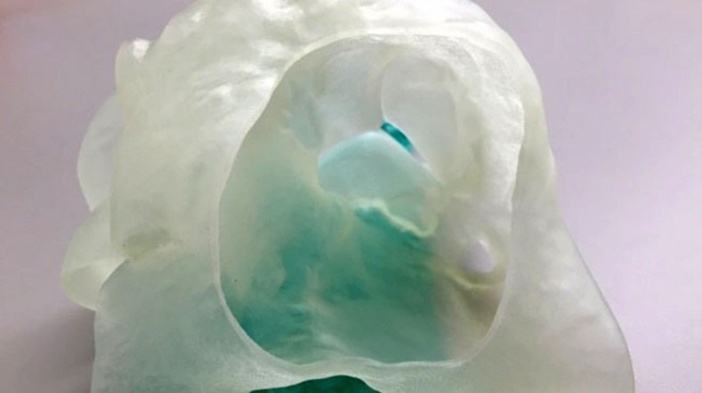
Before a complicated tricuspid valve repair, the Cleveland Clinic 3D-printed a life-size model of a patient's heart. In order to analyze the patient's peculiar anatomy and plan the repair, the medical team utilized the replica.
Even the patient found the model helpful. He gained a better understanding of the physicians' plans after seeing and touching a duplicate of his own heart.
3D Printing Is Not a Replacement for Medical Expertise
3D printing does not make a treatment automatically safer or better. The technology is a tool.
Doctors still need to decide whether a physical model, guide, prosthesis, or other printed device is appropriate. Patient imaging must be processed correctly, the digital model must be accurate, and the final product must meet the required medical standards.
This is why successful hospital programs combine 3D printing with clinical expertise, imaging, engineering, quality control, and medical regulations. The printer is only one part of the system.
Best 3D Design Software
SelfCAD can be a practical 3D design tool for hospitals that need to turn medical scans and digital models into useful 3D-printed objects. Its Bevel, Fillet, Boolean, Stitch & Scoop, and Scale tools give users control over cleaning, editing, combining, and preparing complex models. This can be useful for surgical-planning models, medical training aids, anatomical demonstrations, and custom prototypes.
With its built-in slicer, SelfCAD also lets users prepare finished models for 3D printing without moving to a separate slicing program. For hospitals, this provides a single workspace for editing a model, checking its geometry, and preparing it for fabrication.
Final Thoughts
Hospitals have many reasons to integrate 3D printing. It can turn medical scans into physical models, support surgical planning, create patient-specific guides and devices, provide training tools, and make complex anatomy easier to explain.
Real-world programs at Mayo Clinic, Cleveland Clinic, and Johns Hopkins show that the technology is already being used in clinical and educational workflows. Research has also demonstrated applications ranging from customized casts to patient-specific prostheses.
The most useful applications are not about printing something simply because it is possible. They are about solving a specific clinical problem where a patient-specific physical object can give doctors information, control, or options that a standard product or 2D image cannot provide as easily.